
Hyperspectral Rare Earth Element Mapping
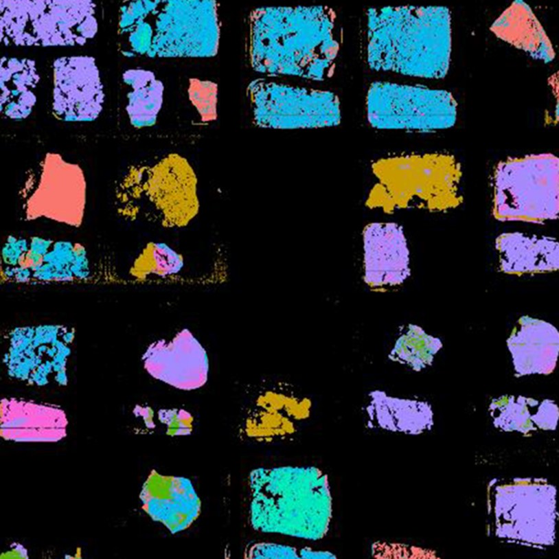
Automatic element and mineral detection in thin sections using hyperspectral transmittance imaging microscopy (HyperTIM).
Automatic element and mineral detection in thin sections using hyperspectral transmittance imaging microscopy (HyperTIM).
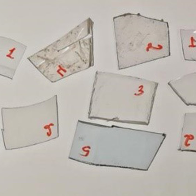
With the advance on net-zero energy technologies, the worldwide demand for Rare Earth Element (REE), e.g., Neodymium (Nd), is predicted to grow. The predicted increase of on- and off-shore wind turbine installations projects a growth in REE metal demand of about 250% to reach the 2-degree scenario through 20501. More efficient detection and classification methods for these REE-metals will be crucial for future exploration and mining. Hyperspectral imaging is already successful to detect REE minerals and elements in outcrops, rock samples, and drill cores using reflectance measurements and allows for the assessment of previously unexplored deposits.2,3,4 The analysis of thin sections plays a crucial role in outcrop characterization but is traditionally achieved using crossed polarized light (XPL), scanning electron microscopy (SEM), electron microprobes (EMP), and point spectrometers (Figure 1).
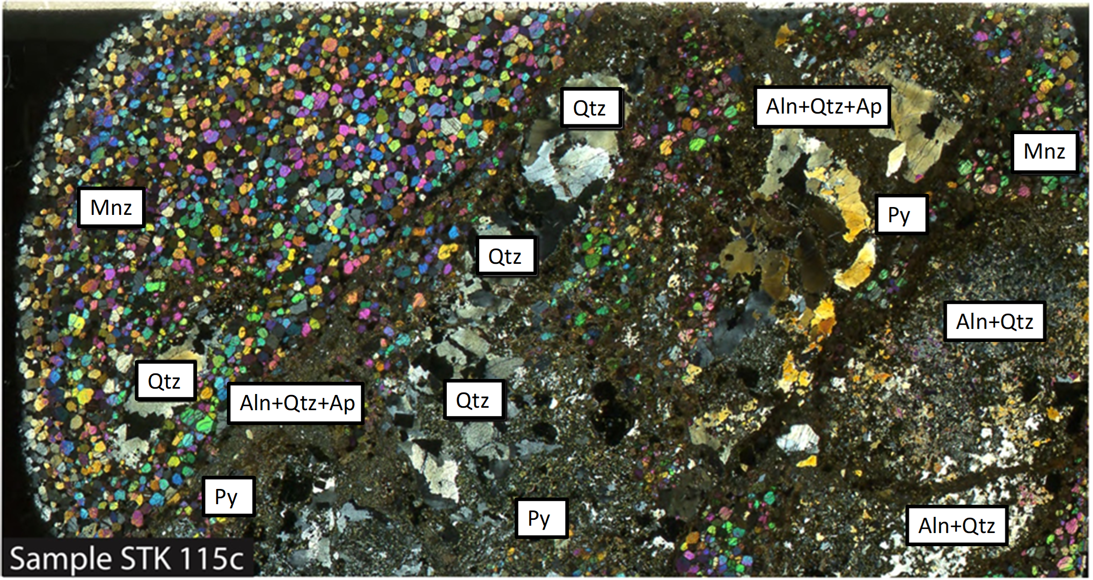
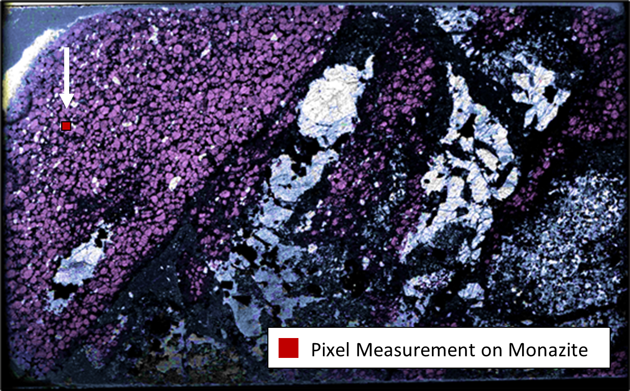
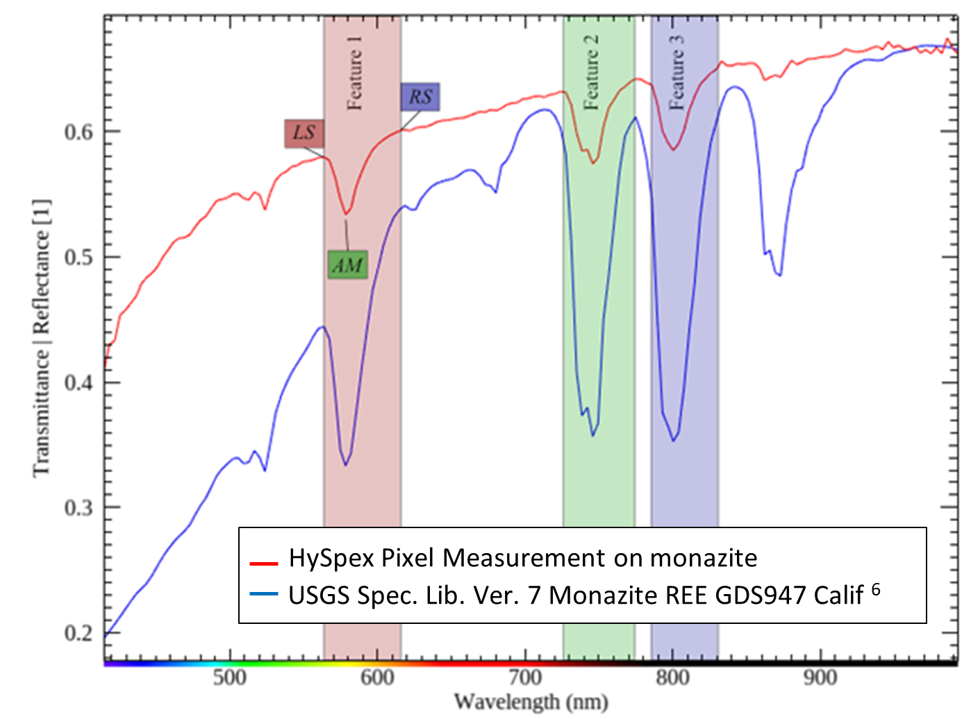
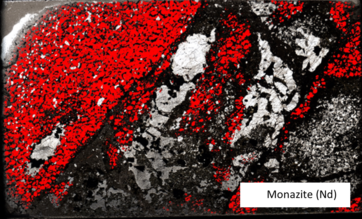
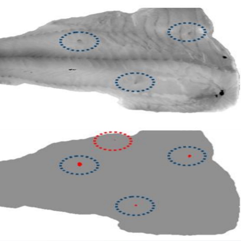
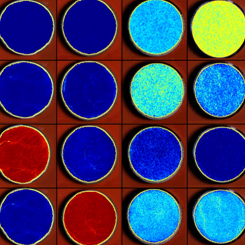
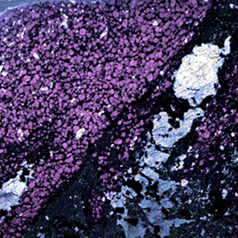
Hyperspectral transmittance imaging microscopy of complete thin sections (HyperTIM) is a novel method for the automated detection of REE minerals.5 HyperTIM uses a HySpex system of the classic series, the VNIR-1600, and a specifically designed sample holder, scanning setup, and microscopic lens. In this example, the method is used on a rock thin section from Steenkampskraal (South Africa), which is analyzed for the REE-bearing mineral monazite ((Ce,Nd,La)PO4), dominated by Nd. Transmittance measurements with the HySpex VNIR camera and a classification algorithm are used to identify and map Nd-bearing monazites in the thin section (Figure 2).
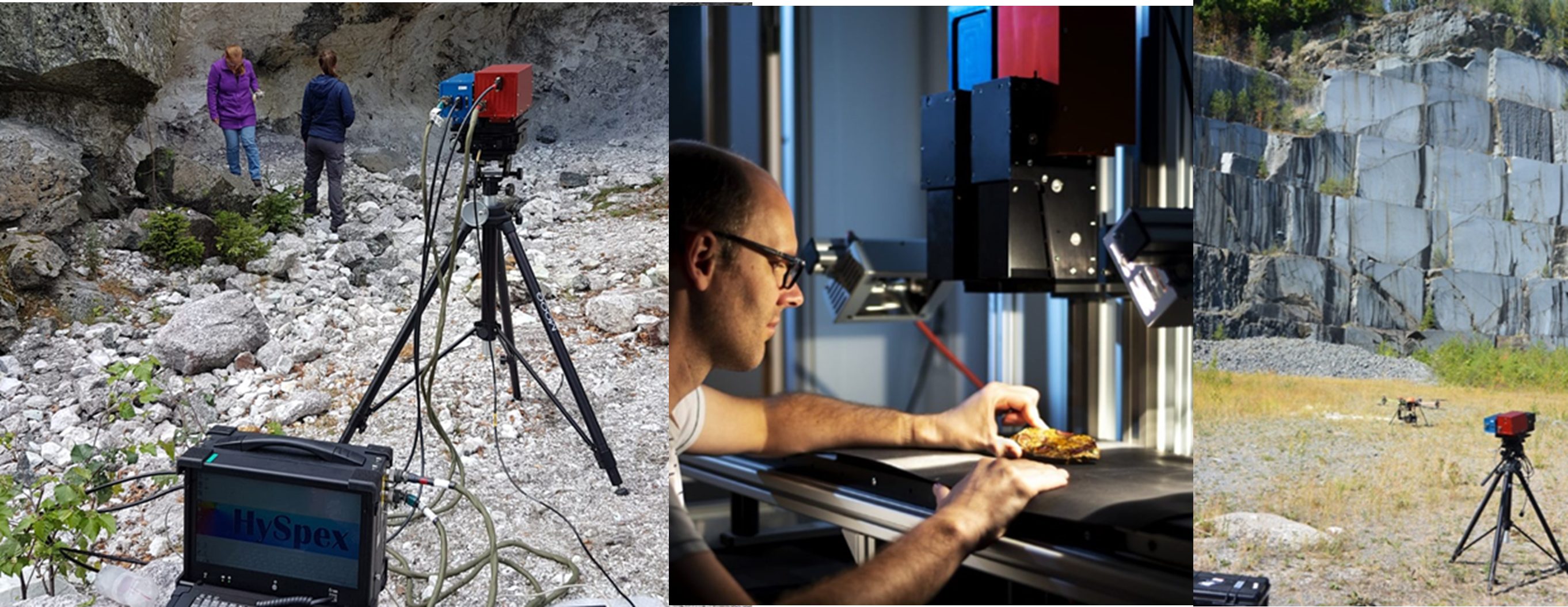
The hyperspectral data in this project was acquired using VNIR and SWIR cameras from the HySpex Classic series. The VNIR-1800 camera covers the 400 – 1000 nm range while the SWIR-384 operates in the 930 – 2500 nm range. The cameras have a spectral resolution of 3.3 and 5.5 nm, respectively.
The cameras are designed to operate in both the laboratory and the field, preserving the spectral fidelity needed for scientific and industrial applications thanks to their low-value optical aberrations, thermal stability and custom lenses for a variety of working distances. The portable field system utilizes a battery-based, rugged data acquisition unit to power and control the cameras as well as the necessary moving stages.
The data analysis and development of HyperTIM was performed within the Rare Earth Element Mapping project REEMAP at the German Research Centre for Geosciences GFZ Potsdam.
This example shows the potential of hyperspectral imaging as a tool for applications in the mining industry. It allows for a precise identification of minerals and materials anywhere from exploration and mining to processing and manufacturing. HySpex offers a varied selection of turn-key solutions for scientific and industrial applications.
Download this application note.

Pharmaceuticals
Methane Detection
Plastic Sorting
Mineral mapping in open pit mines

Asbestos

Explosives
Hyperspectral Imaging for Ore Distinction

Hyperspectral Analysis of Powder Mixtures

Paper Recycling
Rare Earth Elements

Coating thickness

Monument Preservation

Cocoa Beans