
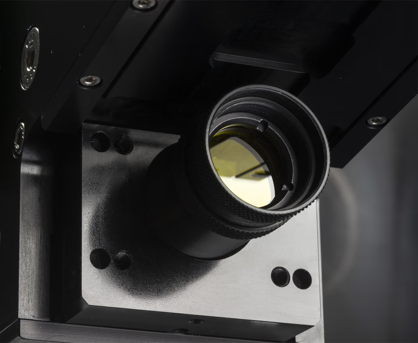

Every hyperspectral image starts with a single instrument: the imaging spectrometer. It is the precision optical device that takes incoming light from a scene, splits it into hundreds of narrow wavelength bands, and records both spatial and spectral information simultaneously. Without it, hyperspectral imaging would not exist.
In modern hyperspectral systems, this instrument is more specifically called a hyperspectral imaging spectrometer — to distinguish it from spectrometers that capture only a handful of bands or that measure single points without spatial information. The scientific method built around using such instruments is known as hyperspectral imaging spectroscopy — and it is what turns raw spectral measurements into mineral maps, vegetation indices, target identifications, and the analytical results that hyperspectral users actually work with.
This article explains what an imaging spectrometer is, how it works, how it compares with other spectrometer types, how it integrates into a complete hyperspectral imaging system, and what separates a research-grade instrument from a casual one. It is the technical foundation that every other piece of hyperspectral content builds on.
What an Imaging Spectrometer Actually Does
An imaging spectrometer captures two pieces of information at every spatial pixel: where the light is coming from in the scene, and how that light is distributed across wavelengths. The result is not a photograph in the conventional sense, but a measurement product where every pixel carries a full spectroscopic signature.
This is a fundamentally different kind of instrument than a normal camera. A camera records three broad colour channels — red, green, and blue — that approximate human vision. An imaging spectrometer records hundreds of narrow channels, typically extending from the visible into the near-infrared and often into the shortwave infrared, capturing how light interacts with materials in ways that no camera and no human eye can detect.
The output of an imaging spectrometer is a hyperspectral image — a three-dimensional data cube where two dimensions are spatial and the third is spectral. Each pixel can be analysed for material composition through its spectral signature, supporting applications from mineral identification to medical research to industrial sorting.
When the spectral sampling is dense enough — hundreds of narrow contiguous bands — and combined with imaging, the instrument is called a hyperspectral imaging spectrometer. The distinction matters because not every spectrometer that produces images is hyperspectral. Some instruments capture only a few bands (multispectral), some sacrifice spectral detail for instantaneous capture (snapshot), and some measure spectra without preserving spatial information (point spectrometers). A hyperspectral imaging spectrometer is specifically the class designed for dense spectral sampling alongside imaging.
The Pushbroom Architecture That Defines Scientific-Grade Imaging Spectrometers
Most scientific-grade hyperspectral imaging spectrometers — including the entire HySpex product range — use a pushbroom design. Understanding this architecture is the key to understanding what makes a modern imaging spectrometer work.
The instrument begins with fore optics: lenses that focus light from a narrow line in the scene onto an entrance slit. This slit defines the spatial line being measured at any moment. Light passing through the slit then enters a collimating optical system that aligns the beam for the next stage.
The collimated light hits a dispersive element — in HySpex spectrometers, a precision transmission grating. The grating separates the incoming light into its constituent wavelengths, fanning out the spectrum across an angular range.
The dispersed light is then focused by focusing optics onto a two-dimensional detector array. On the detector, one dimension corresponds to spatial position along the original line, and the other corresponds to wavelength. The result is a single line of the scene with a full spectrum recorded for every spatial pixel.
To build a complete two-dimensional hyperspectral image, the scene moves relative to the spectrometer — through aircraft motion in airborne acquisitions, conveyor motion in industrial deployments, or controlled stage motion in laboratory work. As successive lines are recorded, they assemble into the three-dimensional data cube that defines a hyperspectral image.
This pushbroom architecture is why scientific-grade hyperspectral spectrometers deliver superior data quality. Each spatial pixel receives an undisturbed full-spectrum measurement; the optical design is optimised for spectral fidelity rather than instantaneous capture; calibration can be characterised precisely across both the spatial and spectral dimensions. The trade-off — that the scene must move relative to the sensor — is acceptable in nearly every demanding scientific or industrial application. Alternatives such as snapshot hyperspectral cameras exist for specialised use cases, but they sacrifice the data quality that serious analytical work depends on.
Where the Imaging Spectrometer Fits Among Other Spectrometer Types
To appreciate what makes an imaging spectrometer distinct, it helps to see the broader spectrometer family.
Point spectrometers measure light from a single location or averaged spot. They are widely used in laboratory chemistry, in field spectroscopy for ground-truthing remote sensing data, and in handheld instruments for spot analysis. They are simpler than imaging spectrometers but provide no spatial information.
Fourier Transform spectrometers (FTIR) use interferometric measurement rather than spatial dispersion. They typically excel at spectral resolution and are common in chemistry and materials analysis. Imaging FTIR systems exist but face engineering challenges that dispersive imaging spectrometers avoid.
Filter-based spectrometers use tunable filters, filter wheels, or fixed bandpass filters to select wavelengths sequentially. They are common in multispectral imaging systems and can be effective when the analyst knows in advance which spectral bands are diagnostic — but they typically lack the dense spectral sampling that hyperspectral analysis requires.
Snapshot spectral imagers capture spatial and limited spectral information in a single exposure, usually through filter arrays integrated with the detector. They trade spectral density and uniformity for instantaneous capture and are useful in scenarios involving rapid motion or short observation windows.
Dispersive imaging spectrometers — the category HySpex builds — combine a slit, grating, and detector array in a pushbroom architecture. Within this category, several optical layouts are used. Czerny-Turner spectrometers are a classic design with separate collimating and focusing mirrors; they are well-understood and widely used. Offner spectrometers employ a single curved grating in a compact relay arrangement and are common in space-based remote sensing payloads. Dyson spectrometers use a single grating with immersion optics for very compact designs that excel in size-constrained applications. Each layout has trade-offs in compactness, optical performance, manufacturing complexity, and stray light behaviour.
HySpex hyperspectral imaging spectrometers are built around optical designs developed in-house, optimised for the specific combination of properties that demanding hyperspectral applications require: spectral fidelity, spatial sharpness, calibration stability over years of operation, and integration into platforms ranging from laboratory benches to UAVs to satellites.
What Separates a Research-Grade Imaging Spectrometer from a Casual One
The most important thing to understand about imaging spectrometers is that two instruments with identical headline specifications can deliver very different data quality. The differences live in details that rarely appear on spec sheets.
Smile and keystone are small geometric distortions in the spectral and spatial dimensions, respectively. A spectrometer with significant smile records slightly different wavelengths at different positions along the spatial line, undermining the spectral integrity that analytical workflows depend on. Keystone distortions cause spatial misregistration across wavelengths, with similar effects on data quality.
Stray light is unwanted light that reaches the detector through paths other than the intended optical design — internal reflections, scattering from optical surfaces, or light leakage from adjacent wavelengths. It contaminates measurements particularly where the analytical signal is weak, and it can be a significant differentiator between instruments that appear similar on a spec sheet.
Polarisation sensitivity can affect reflectance measurements in unexpected ways, especially in airborne and field deployments where surface and atmospheric polarisation effects vary across the scene. Well-designed scientific-grade spectrometers minimise polarisation sensitivity through careful optical design.
Calibration stability over time is what allows data acquired today to be compared with data acquired years from now — across temperature changes, mechanical handling, and operational wear. This is the foundation of any longitudinal analytical programme.
These quality factors are covered in depth in the HySpex Key Quality Parameters resources. For users evaluating imaging spectrometers, the practical implication is that comparing nominal specifications between instruments is a starting point — but the real differentiator is the engineering discipline that determines whether the instrument actually delivers the data quality its specifications promise.
For applications where measurements need to be defensible, repeatable, and comparable across instruments and over time, HySpex provides traceable calibration to standards such as NIST and PTB — particularly in the HySpex Baldur industrial series. This level of metrological discipline is what distinguishes scientific-grade imaging spectrometers from less rigorous alternatives.
Spectral Range and What It Reveals
Hyperspectral imaging spectrometers typically operate across one or more spectral regions, each of which reveals different material properties.
The visible and near-infrared (VNIR) region, roughly 400–1000 nm, captures information about vegetation health, water content, pigments, and certain mineral properties. Most general-purpose hyperspectral applications use VNIR coverage as a baseline.
The shortwave infrared (SWIR) region, roughly 1000–2500 nm, is particularly powerful for mineralogy, chemistry, and material identification. Many minerals, plastics, pharmaceuticals, and organic compounds have diagnostic absorption features in the SWIR that allow them to be identified spectrally.
Combined VNIR and SWIR systems — such as the HySpex VS-1200 used in airborne deployments — provide the broadest analytical capability by covering both regions in a single integrated instrument.
In specialised cases, hyperspectral imaging spectrometers extend into the mid-wave and long-wave infrared for thermal and emission-based measurements. These regions support applications such as thermal characterisation and certain industrial process monitoring. (For methane detection specifically, HySpex has developed a satellite-based hyperspectral camera operating in the SWIR through ESA's InCubed programme.)
The spectral range needed depends entirely on the application. Mining and mineral exploration typically prioritises SWIR; vegetation monitoring usually emphasises VNIR with red-edge sensitivity; medical and biomedical research often uses VNIR with extended near-infrared coverage. Selecting the right spectral coverage for the application is one of the foundational decisions when specifying an imaging spectrometer.
Imaging Spectroscopy as a Scientific Discipline
While imaging spectrometer and hyperspectral imaging spectrometer refer to the instrument, imaging spectroscopy — and its more specific variant hyperspectral imaging spectroscopy — refers to the broader scientific discipline of using such instruments to extract material information from spectroscopic measurements organised in space. The terms are often used interchangeably, particularly in academic and remote sensing literature, where "imaging spectroscopy" tends to be preferred over "hyperspectral imaging" in some scientific traditions.
The discipline rests on three foundational principles that have matured over the past several decades.
First, the spectral signature of a material — how it reflects, absorbs, or emits light across wavelengths — is characteristic of its chemical and physical structure. Different minerals, plant species, polymers, tissues, and chemical compounds have distinctive signatures that can be measured and recognised. Imaging spectroscopy is the systematic exploitation of this principle across entire scenes rather than at single points.
Second, calibration and traceability are essential. A spectral signature is only meaningful if measurements can be tied to physical units and compared against reference standards. Without calibration, an apparent spectral feature might be an instrument artifact; with proper calibration, it can be matched against established spectral libraries such as those maintained by USGS or ASTER. Our overview of hyperspectral image data covers this foundation in more depth.
Third, the analytical methods for interpreting imaging spectroscopy data have grown into a sophisticated toolkit. Classical methods include spectral library matching, spectral angle mapping (SAM), and continuum-removed feature analysis. Modern methods extend into dimensionality reduction (PCA, MNF), spectral unmixing for sub-pixel material abundance, and increasingly machine learning approaches that combine spatial and spectral pattern recognition. Our overviews of hyperspectral image processing and spectral image analysis cover the analytical layer in depth.
For organisations approaching the field, the practical implication is that hyperspectral imaging spectroscopy is no longer experimental. It is a mature discipline supported by well-understood instruments, rigorous calibration practices, and a substantial analytical toolkit. The remaining question for most users is not whether the technology works, but which instrument, deployment configuration, and analytical workflow fit their specific application.
Applications Across Industries
Imaging spectrometers are used in a remarkably wide range of applications, with each industry shaping the instrument requirements in different ways:
- Industrial sorting and quality control — using imaging spectrometers on production lines for materials identification, contamination detection, and process monitoring
- Geological and mineral analysis — applying imaging spectroscopy to drill core scanning, mine face mapping, and mineral exploration
- Vegetation and agricultural monitoring — using imaging spectrometers for crop health, species classification, and yield assessment
- Laboratory measurements and scientific research — supported by Laboratory Systems built around imaging spectrometers with controlled illumination and scanning stages
- Airborne and satellite remote sensing — where imaging spectrometers integrated with navigation and atmospheric correction support wide-area mapping
- Cultural heritage and art conservation — where imaging spectrometers reveal pigment composition and hidden layers in artworks
- Defense, surveillance, and ISR — where imaging spectrometers support target detection and material identification
- Medical and biomedical research — where imaging spectrometers enable tissue oxygenation and composition measurements
Each application places different demands on the imaging spectrometer. A spectrometer optimised for industrial sorting needs robust performance at high data rates with reliable real-time integration. One designed for airborne surveys needs calibration stability across multi-hour flights and integration with navigation systems. One built for laboratory research prioritises spectral fidelity and analytical flexibility. The same underlying technology serves all of these, but the specific configurations and supporting components differ substantially.
From Standalone Instrument to Integrated System
A modern imaging spectrometer is rarely deployed in isolation. It sits at the heart of an integrated hyperspectral imaging system that includes the spectrometer itself, fore optics matched to the application, acquisition electronics, calibration components, mounting hardware appropriate to the deployment environment, and the software stack that turns raw data into useful results.
HySpex organises its complete systems around three camera families. The HySpex Classic series provides scientific-grade general-purpose imaging spectrometers for laboratory, field, and airborne use. The HySpex Mjolnir series packages the technology in compact form factors for UAV deployment and field-portable applications. The HySpex Baldur series is purpose-built for industrial deployment with traceable calibration and integration features for production environments.
The software layer that makes these instruments practically useful includes the Prediktera Software Suite for analysis and modelling, ATCOR-4 for atmospheric correction in airborne applications, and PARGE for geometric correction. For applications that require decisions during acquisition rather than after, HySpex Bifrost performs real-time georeferencing, atmospheric correction, and application-specific modelling on the fly.
Machine learning and AI are increasingly integrated into hyperspectral workflows, with classical spectral methods now complemented by deep learning approaches that capture combined spatial-spectral patterns. The combination of robust optical instrumentation, mature analytical software, and modern computational methods is what turns a hyperspectral imaging spectrometer from an interesting capability into an operational tool.
The Engineering Heritage Behind Scientific-Grade Imaging Spectrometers
The performance of an imaging spectrometer is shaped as much by the engineering culture that built it as by the components inside it. HySpex traces its roots to Norsk Elektro Optikk, founded in 1985 by researchers from the Norwegian Defence Research Establishment (FFI) working on electro-optical instrumentation. The first HySpex hyperspectral cameras went into production in 2003, and the product line has continued to evolve over more than two decades of operational use across science, industry, defence, and space.
This continuity matters operationally. Long-running scientific programmes depend on imaging spectrometers that behave consistently over years of work, on the ability to compare data acquired with different but related instruments, and on the availability of calibration services and engineering support over multi-year deployment cycles. HySpex reports that none of its hyperspectral instruments have reached end of life — a continuity that underpins the long-term analytical investments users build around their hyperspectral capability.
Summary: The Imaging Spectrometer at the Heart of Hyperspectral Imaging
A hyperspectral imaging spectrometer is the core optical instrument that makes hyperspectral imaging possible. By capturing spatial and spectral information simultaneously across hundreds of narrow wavelength bands, it produces the data that hyperspectral imaging spectroscopy workflows turn into mineral maps, target identifications, vegetation indices, quality control decisions, and the analytical results that define real-world hyperspectral applications.
The instrument exists in many forms — from compact UAV cameras to airborne survey instruments to industrial production line scanners to laboratory research platforms — but the underlying principles are shared. Optical quality, calibration discipline, integration engineering, and analytical software all contribute to what makes the difference between a hyperspectral imaging spectrometer that delivers useful data and one that does not.
Selecting an Imaging Spectrometer for Your Application
Choosing the right hyperspectral imaging spectrometer involves more than reviewing top-level specifications. Optical performance, spectral stability, calibration strategy, deployment environment, and integration with your analytical workflow all influence what the instrument can actually deliver in practice — and the right choice depends on whether your application emphasises the imaging spectroscopy side (analytical depth, calibration traceability, library matching) or the imaging system side (deployment flexibility, real-time integration, operational robustness).
HySpex works with research institutions, industrial users, system integrators, and operational programmes to configure imaging spectrometers and complete hyperspectral systems for laboratory, field, airborne, UAV, and industrial environments. If you are evaluating an imaging spectrometer for a specific application, our team can provide technical guidance on system design, performance trade-offs, and integration paths. Feel free to contact us for more information.
FAQ – Imaging Spectrometers and Imaging Spectroscopy
What is an imaging spectrometer?
An imaging spectrometer is an optical instrument that captures both spatial and spectral information from a scene. Unlike a conventional camera that records three broad colour channels, an imaging spectrometer records hundreds of narrow wavelength bands at every spatial pixel, producing a three-dimensional data cube suitable for material identification, classification, and quantitative analysis.
What is a hyperspectral imaging spectrometer?
A hyperspectral imaging spectrometer is an imaging spectrometer designed specifically for hyperspectral measurement — capturing many narrow contiguous wavelength bands at every spatial point in a scene. The term emphasises both the imaging capability (preserving spatial information) and the hyperspectral character (dense spectral sampling rather than discrete band selection). HySpex hyperspectral imaging spectrometers are built around dispersive pushbroom architectures optimised for spectral fidelity, calibration stability, and integration into complete hyperspectral imaging systems.
What is imaging spectroscopy?
Imaging spectroscopy is the scientific discipline of using imaging spectrometers to measure spectral signatures across entire scenes. It is closely related to — and often used as a synonym for — hyperspectral imaging, particularly in academic and remote sensing contexts. The discipline combines instrument design (imaging spectrometers) with calibration methodology and analytical techniques to support material identification, classification, and quantitative measurement.
Is hyperspectral imaging spectroscopy the same as hyperspectral imaging?
The terms are essentially synonymous in practice. Hyperspectral imaging spectroscopy emphasises the spectroscopic measurement aspect of the technology — the fact that each pixel carries a full spectrum analysable for material composition. Hyperspectral imaging is the more common umbrella term that includes both the instrument (the hyperspectral imaging spectrometer) and the resulting imagery and workflow. Academic and remote sensing literature often prefers "imaging spectroscopy" or "hyperspectral imaging spectroscopy"; commercial and industrial contexts often prefer "hyperspectral imaging".
What are the main types of spectrometer?
The main spectrometer types include point spectrometers (measuring spectra from a single location), Fourier Transform spectrometers (using interferometric measurement), filter-based spectrometers (using fixed or tunable filters), snapshot spectral imagers (capturing limited spectral information instantaneously), and dispersive imaging spectrometers (using slit, grating, and detector array to capture spatial and spectral information). Imaging spectrometers used in scientific-grade hyperspectral systems are typically dispersive designs based on the pushbroom architecture, with internal layouts including Czerny-Turner, Offner, and Dyson variants.
What is the difference between an imaging spectrometer and a hyperspectral camera?
In most modern systems, the two terms refer to closely related concepts. A hyperspectral camera is typically built around an imaging spectrometer, which is the optical instrument responsible for separating light into many spectral bands while preserving spatial information. The camera adds the housing, electronics, fore optics, and integration features that make the spectrometer usable in a specific deployment.
What spectral range do imaging spectrometers cover?
Imaging spectrometers typically cover one or more spectral regions: visible and near-infrared (VNIR, roughly 400–1000 nm), shortwave infrared (SWIR, roughly 1000–2500 nm), or combined VNIR+SWIR systems for the broadest analytical coverage. Specialised instruments extend into mid-wave and long-wave infrared regions. The right spectral range depends on the application — mining typically uses SWIR for mineralogy; vegetation monitoring often uses VNIR; many research applications benefit from combined VNIR+SWIR coverage.
How is data quality maintained in imaging spectrometers?
Data quality depends on optical design (minimising smile, keystone, stray light, and polarisation sensitivity), calibration discipline (traceable to standards such as NIST and PTB), and stable mechanical and thermal design. HySpex's Key Quality Parameters resources cover the factors that distinguish research-grade instruments from casual ones.
Can imaging spectrometers be used outside the laboratory?
Yes. Modern imaging spectrometers are integrated into systems for industrial production lines, airborne platforms, UAVs, and satellites. The HySpex turnkey product range covers laboratory, field, airborne, UAV, and industrial deployment configurations, with custom solutions developed for applications outside these standard configurations.